Full-Stack Developer & Scientific Software Engineer @ Biocomputing Unit

We have a Full-Stack Developer & Scientific Software Engineer position open at the Biocomputing Unit. Get to know it in this post!

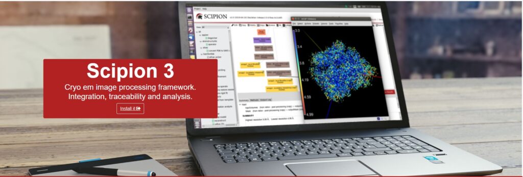
About Scipion: Scipion is an open-source, workflow-oriented software framework used primarily in Cryo-Electron Microscopy (cryoEM). It integrates various scientific software packages into a unified interface, allowing researchers to execute, track, and share complex image-processing pipelines. Its goal is to ensure reproducibility and provide a seamless experience for processing high-resolution biological data.

About ScipionWeb: ScipionWeb is the modern web-based evolution of this framework. It provides a platform to manage scientific projects and workflows: from project creation and protocol execution to high-level visualization and productivity tools.

Stack: React + TypeScript (Frontend) and FastAPI + Python (Backend).

Your Tasks & Evolution: In this role, your responsibilities will evolve as you gain mastery over the framework:

Short to Medium Term:

  • 50% Web Development: Full-stack development of the ScipionWeb interface (UI + API + Integration).
  • 50% Scipion Core & Domains: Diving into the backend logic, the Scipion core architecture, and specific scientific domains like SPA (Single Particle Analysis) and Tomography.

Medium to Long Term:

  • Focus on the Scipion Core and expansion into new scientific domains.
  • Maintain and update the ScipionWeb ecosystem to ensure long-term stability.

    Requirements

    Academic & Language

    • Degree: BSc/MSc in Physics, Mathematics, or Software Engineering.
    • English: B2 level or equivalent (ability to work in an international environment).

    Technical Essentials

    • Python: 2+ years of experience with Core Python (Standard Library).
    • Linux: Solid basic knowledge (Ubuntu is our preferred distribution).
    • Version Control: GitHub (PRs, collaborative workflows).
    • DevOps/Distribution: Experience with software distribution (PyPI, Conda) and CI/CD (GitHub Actions).

    Stack & Desired Skills

    The Tools We Use:

    • Frontend: React Router, React Query, MUI, ReactFlow (for diagrams).
    • Backend: FastAPI, Pydantic, service-oriented layers.
    • Integration: Async workflows (jobs/queues), schema-based forms, and data visualization.

    Bonus Points (Desired Skills):

    • Knowledge of Image Processing or cryoEM.
    • Experience with 3D visualization (three/react-three-fiber or vtk.js).
    • Experience with Docker and Celery.
    • A strong eye for API design and complex UI (editors, dashboards, large-scale data).

    What We Value

    We are looking for someone with a scientific mindset—someone who is not only a great coder but is also curious about how pipelines and HPC (High-Performance Computing) environments work. You should enjoy solving complex logic puzzles and building tools that help scientists discover the building blocks of life.

    Applying

    Are you interested in our Systems Manager job position? Send your CV and a letter of interest to blanca@cnb.csic.es.

    2 Open Positions: Software Developer at Biocomputing Unit (BCU), Instruct Image Processing Center (I2PC), Madrid

    Description: We are looking for two candidates with a BSc/MSc in Computer Science, Mathematics, Engineering, Physics or equivalent with software development skills. The candidate will be involved in European and National projects related to cloud and open data technologies, being part of a team in charge of developing data management software for Instruct facilities.

    Thus, the candidate will participate in projects that will shape and influence strategies for making research data generated across the Instruct-ERIC infrastructure FAIR (Findable, Accessible, Interoperable, Reusable). This data transformation will involve considerations of the storage, metadata description and deposition of large datasets for use and reuse including artificial intelligence. These data will come primarily from structural biology but can include data from other diverse research domains.

    Background: INSTRUCT ERIC is the European Strategic Initiative in the area of Integrative Structural Biology. It is organized as a distributed infrastructure with “Instruct Centers” and the BCU hosts the “INSTRUCT Image Processing Center (I2PC)”.

     BCU is well known in the area of 3D-EM, with over 200 publications in the area of Electron Microscopy and large contributions to open source scientific software, like Scipion, Xmipp and 3DBionotes.

    For further information go to INSTRUCT Image Processing Center or  Biocomp web sites.

    Location: Madrid 

    Required Skills:

    • BSc / MSc in Computer Science, Mathematics, Engineering, Physics or equivalent
    • Programming experience with Python
    • Experience in Linux
    • Medium level of proficiency in written and spoken English

    The following skills are considered a “plus”, but are not essential:

    • Experience of the full lifecycle of software architecture, design, and implementation
    • Experience developing web and desktop Python apps
    • Knowledge and some degree of experience using relational and/or noSQL databases.
    • Experience in assisting open source software development teams (GitHub, documentation, user support, collaboration tools…).
    • Interest or experience in scientific data management
    • Interest or experience in biological sciences or bioinformatics
    • Experience in Virtualization: Docker

    Interested candidates should send their CV’s and letter of interest to blanca@cnb.csic.es

    An asymmetric structure of the Leptospira´s flagellar filament

    Date and venue:  2024, February 22th al 10.00 CET.
    B6 room- CNB/CSIC, Madrid

    Summary Description:
    Leptospira species, as all spirochetes, possess specially adapted endoflagella confined within the periplasm. Endoflagella are essential for the fast corkscrew-like swimming and pathogenicity of leptospires. However, little is known about the composition, function, and molecular architecture of these endoflagellar filaments. Through a combination of bacterial genetics, protein crystallography and cryo-electron microscopy/tomography, significant progress has been made in determining the 3D structures of complete Leptospira flagellar filament assemblies, revealing an asymmetric organization, comprising at least 14 different types of proteins.
     

    Present by: Nicole Larrieux from Unidad de Cristalografía de Proteínas (Laboratorio de Microbiología Molecular y Estructural at IPM)

    For more information, please contact Oscar Saiz at oscar.saiz@cnb.csic.es

    Structural studies on cyanide degrading nitrilases

    Date and venue:  2024, February 15th al 10.00 CET.
    B6 room – CNB/CSIC

    Summary Description:

    Cyanide is widely used in industries due to its high affinity for metals. This characteristic is at the same time responsible for the potent toxicity of this compound. Thus, industries have to reduce the cyanide concentration from wastewater before its final disposal. Methods to achieve this goal exists but they can be improved by increasing our knowledge in the action of enzymes involved in the process. We are studying cyanide hydratases and dihydratases mainly at the structural level with the aim of understanding its basic features for functioning and to propose methods to improve their characteristics. We have solved the structure of two of them, and now we are improving the resolution of our maps, characterizing other homologs and proposing changes to improve them.

    Specifically, we will prepare grids of the different versions of CynD at different pHs. Then, we will screen the grids and collect datasets of the best preparations. At the same time, we will process the previously collected datasets and the newly collected datasets with assistance of expertise of the Jose Maria Carazo team, using more sophisticated software and methodologies characterize conformational heterogeneity and dynamics in cryoEM datasets. In this way, we will gain experience in these state-of-the-art methods.

    Present by: Santiago Justo Arévalo (Instituto de Química- UPS)


    For more information and to request assistance, please contact Oscar Saiz at oscar.saiz@cnb.csic.es

    Structural Biology 2.0 : computational tools and X-ray diffraction integrated to solve and understand 3D protein structures

    Date and venue:  2025, from April 21 to 30. Institut Pasteur de Montevideo (Uruguay).
    All invited speakers/tutors and students from abroad will be lodged in the same hotel.
    Webpage under construction: stay tuned for upcoming information!

    Summary Description: The workshop will cover theory and hands-on practical work, the latter including tutorials and the students’ own data processing. 
    The borders among different approaches to do research in modern Structural Biology, are quickly being blurred. Experimental data are increasingly combined with computational methods in each and every step of the way within real life Biology projects. This advanced, hands-on workshop aims at filling in the gap, avoiding separate chapters for different techniques and instead mimicking real-life research, with these different components interacting all along the process. 
    World experts in X-ray crystallography and Computational approaches (3D structure prediction, Molecular Dynamics simulations) will be invited to the staff of teachers/tutors.
    The workshop is conceived for 20-25 attendees: advanced graduate students, postdocs and young researchers working in Structural Biology, Protein Science and related areas.
    Scholarships will be available to cover students’ costs.
    Students that bring their own data (X-ray diffraction data, and/or actual novel protein structures to analyze) will be given priority at the time of appraisal.
     

    Organization committee: Nicole Larrieux, Natalia Lisa, Matias Machado, Martin Graña, Felipe Trajtenberg, Ari Zeida, Alejandro Buschiazzo.

    I2PC Course on Single Particle Analysis by CryoEM. Madrid, 20 – 22 March, 2024

    Venue

    National Center for Biotechnology (CNB), Madrid, Spain
    Madrid, March 20 – 22, 2024
    How to find us: https://idospece.i2pc.margaro.eus/contact-us/

    Overall aims and course outline

    Cryo-Electron Microscopy has established as a mature technique to determine the three-dimensional structure of biological macromolecules. It is currently reaching atomic resolution and it is capable of accessing to the structure of conformationally heterogeneous samples, as well as macromolecular complexes. It is complementary to other structural techniques like X-ray crystallography and Nuclear Magnetic Resonance and at the moment it is the most rapidly growing technique in atomic structure depositions at public databases like PDB.

    The aim of the course is to give an overall overview of the image processing in single particle analysis (SPA). The I2PC has a long tradition of organizing courses of image processing and atomic modelling, with Instruct courses that have been heavily oversubscribed and had to be repeated twice in the same year to be able to attend its demand.

    With the experience gained in the organization of virtual and in-person courses, we plan to organize this course in an in-person modality. 20 people will be able to physically attend.

    The course is aimed at researchers of all levels wanting to adopt electron microscopy as one of the tools at their disposal for elucidating biological structures.

    For more information see:

    I2PC Course on Single Particle Analysis by CryoEM. Madrid, 20 – 22 March, 2024 – I2PC – Instruct Image Processing Center

    I2PC – Instruct-RI Hubs hybrid course on Electron Tomography and Subtomogram Averaging. Madrid, December 10 – 13, 2024

    VENUE

    Hybrid course.
    December 10 – 13, 2024 from 16:00 to 21:30 CET.
    You do not need to install any special software to connect. A modern browser should be enough.

    Overall aims and course outline

    By using image processing algorithms, it becomes possible to pinpoint the macromolecules of interest within the electron tomograms. Subsequently, researchers can extract small subtomograms from these locations and identify similar particles, facilitating their averaging and the recognition of diverse conformations. This process, referred to as subtomogram averaging, has played a pivotal role in elucidating an increasing number of macromolecular structures, achieving resolutions ranging from 20 to 3 A.

    The course is designed to offer a comprehensive overview of the whole image processing workflow in Electron Tomography (ET). This includes tasks such as aligning the tilt series, reconstructing 3D tomograms, identifying particles, performing subtomogram averaging or even to perform a tomogram segmentation, all inside Scipion integration framework.  

    With the experience gained in the organisation of virtual and in-person courses, we intend to organise this course in a hybrid format. We will be able to accommodate up to 20 individuals for in-person attendance. However, other 20 participants will attend remotely. In total, we can host 40 attendees. 

    The course is aimed at researchers of all levels wanting to adopt electron microscopy as one of the tools at their disposal for elucidating biological structures

    For more information see:

    I2PC – Instruct-RI Hubs hybrid course on Electron Tomography and Subtomogram Averaging. Madrid, December 10 – 13, 2024 – I2PC – Instruct Image Processing Center

    Fundamentals of light microscopy and image processing

    Date and venue: 2023, 18-29 of September at ITQB-NOVA. Will transmit lectures over Zoom: https://videoconf-colibri.zoom.us/j/95295678533

    Summary Description: The course “Fundamentals of light microscopy and image processing” will focus on the application of fluorescence microscopy in molecular and cellular microbiology and infection biology. This course will pass on basic concepts and practical training in conventional epifluorescence and confocal microscopy, super-resolution and electron microscopy. Trainees will learn principles of sample preparation and advanced applications in fluorescence microscopy (FRET, FLIM and FRAP). Through a combination of theoretical presentations and interactive coding sessions, trainees will learn methods of image analysis and machine learning. These concepts and tools will be applied in small group projects in interaction with the trainers.

    Tuesday 19th:

    • 10:30 – 11:30. Part I. Introduction to the basic principles of light microscopy. Davide Accardi – Champalimaud Foundation, Portugal
    • 12:00 – 13:00. Part II. Introduction to the basic principles of light microscopy. Davide Accardi – Champalimaud Foundation, Portugal

    Wednesday 20th

    • 9h00 – 10h00. Introduction to the principles of sample preparation in fluorescence microscopy. Hannah Heil – IGC, Portugal.
    • 10h30 – 12h00. Basic principles of widefield and widefield Super Resolution microscopy. Zach Hensel – ITQB-NOVA, Portugal.
      14h00 – 15h00.  Introduction to expansion microscopy.  Mario Del Rosario – IGC, Portugal.

    Friday 22nd

    • 9h00 – 10h00. Part I – Basic principles of confocal microscopy.  José Rino – IMM, Portugal.
    • 10h30 – 12h00. Part II – The F techniques: FRAP, FRET and FLIM.  José Rino – IMM, Portugal.
    • 12h00 – 13h00. Basics of Electron Microscopy. Ana Sousa – IGC, Portugal.

    For more information see:

    https://www.itqb.unl.pt/events/short-course-fundamentals-of-light-microscopy-and-image-processing

    Position Open in image processing for Cryo-Electron Microscopy at BCU

    The Biocomputing Unit (BCU) of the Natl. Center of Biotechnology is looking for a candidate with a background in Engineering, Physics, Mathematics or any other data analysis related field. The work is about the development of image processing in Cryo-Electron Microscopy (CryoEM) with a special emphasis on signal processing and efficient implementations. Our laboratory is located at the National Center of Biotechnology in Madrid, a reference institute in Spain in cryo-EM equipped with two microscopes with direct detector (Talos Arctica and CryoARM 300) and a FIB-SEM microscope, providing a very immersive and interdisciplinary environment. Additionally, our group is a world reference in this domain, and the work will be performed in conjunction with an American/Canadian drug discovery company. The goal of the project is to construct the most efficient 3D reconstruction algorithm available for CryoEM.

    Recent publications:

    FSC-Q: a CryoEM map-to-atomic model quality validation based on the local Fourier shell correlation. Ramírez-Aportela E, Maluenda D, Fonseca YC, Conesa P, Marabini R, Heymann JB, Carazo JM, Sorzano COS. Nat Commun. 12(1):42. 2021.
    DOI: https://doi.org/10.1038/s41467-020-20295-w

    Measuring local-directional resolution and local anisotropy in cryo-EM maps. Vilas JL, Tagare HD, Vargas J, Carazo JM, Sorzano COS. Nat Commun. 11(1):55. 2020.
    DOI: https://doi.org/10.1038/s41467-019-13742-w

    Continuous flexibility analysis of SARS-CoV-2 spike prefusion structures. Melero R, Sorzano COS, Foster B, Vilas JL, Martínez M, Marabini R, Ramírez-Aportela E, Sanchez-Garcia R, Herreros D, Del Caño L, Losana P, Fonseca-Reyna YC, Conesa P, Wrapp D, Chacon P, McLellan JS, Tagare HD, Carazo JM. IUCrJ. 7(Pt 6):1059-69. 2020.
    DOI: https://doi.org/10.1107/S2052252520012725

    Measurement of local resolution in electron tomography. Vilas JL, Oton J, Messaoudi C, Melero R, Conesa P, Ramirez-Aportela E, Mota J, Martinez M, Jimenez A, Marabini R, Carazo JM, Vargas J, Sorzano COS. J Struct Biol X. 4:100016. 2019
    DOI: https://doi.org/10.1016/j.yjsbx.2019.100016

    DeepRes: a new deep-learning- and aspect-based local resolution method for electron-microscopy maps. Ramírez-Aportela E, Mota J, Conesa P, Carazo JM, Sorzano COS. IUCrJ. 6(6): 1054 – 1063. 2019
    DOI: https://doi.org/10.1107/S2052252519011692

    Interested candidates should send their CV’s and letter of interest to: blanca@cnb.csic.es